The reasons for this operation are usually polyps in cats and chronic ear inflammation in dogs. Tumours are occasionally the reason for the surgical procedure.
In cats, the approach is usually from the front, i.e., from the underside of the head as the middle ear is relatively superficial here and is very readily accessible. The middle ear lies much deeper in dogs. Here it depends on the individual case whether it is accessed from below or from the side (laterally). With the lateral approach, the external auditory canal is also removed. This is called ablation of the auditory canal with lateral bulla osteotomy and this approach is chosen when the disease also affects the auditory canal.
The bony boundary of the middle ear is opened and all inflamed or tumour tissue and/or secretion is removed.
Horner’s syndrome is a very common complication after bulla osteotomy in cats. Very fine nerve fibres that run through the middle ear in cats are often irritated during the operation so that nictitating membrane protrusion, a narrow pupil and drooping upper lid occur subsequently in the eye on the same side. Horner‘s syndrome usually resolves spontaneously within two weeks.
With the lateral approach, irritation of the facial nerve can occur as a complication. This happens more often in cats than in dogs because the nerves are thinner and more fragile overall. In this case, the eyelid no longer closes correctly and it is necessary postoperatively to moisten the eye several times a day with eyedrops. In this case, too, nerve function usually returns to normal within a few weeks.
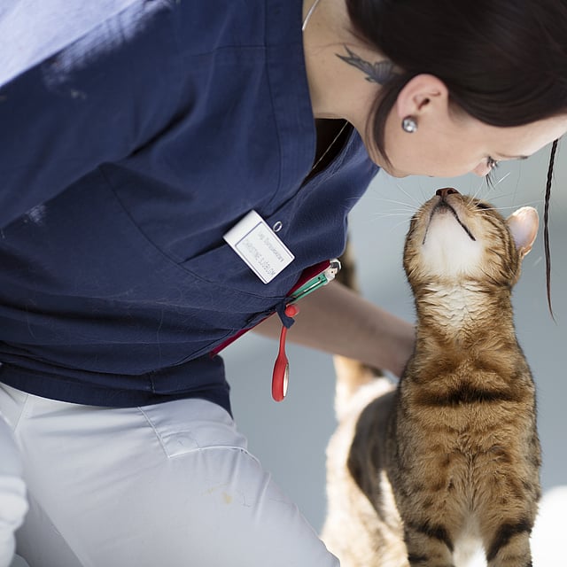
© AniCura, Dr. Riccarda Schünemann