In veterinary medicine dogs must be given a light anaesthetic for a CT scan. The examination lasts only a few minutes, however, so it is possible even for dogs with an increased anaesthetic risk, such as old, sick or injured dogs.
Due to the short duration of the anaesthetic, CT is also very suitable for diagnostic investigations prior to necessary surgical procedures.
It is very useful for operation planning as it shows complex fractures accurately. Even hairline cracks not identifiable on an ordinary X-ray (e.g., of the skull) become visible.
In trauma diagnostics, it can show if there are bone fractures and/or haemorrhage (e.g., with head injury) or internal injuries. Since computed tomography can be performed in a very short time, it provides a rapid and comprehensive overview especially in polytrauma animals (i.e., when many organs are injured) of all the patient‘s injuries without stressing him with unnecessarily prolonged anaesthesia.
How is a CT scan done?
A CT scan can only be performed in dogs under a brief anaesthetic. Following premedication, this generally uses inhalational anaesthesia with isoflurane and oxygen. A contrast agent is often injected through a vein in addition.
The scan takes a few minutes.
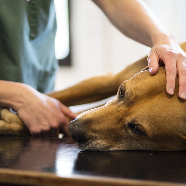
At the end of the examination, the patient is monitored and the drip infusion continues until he has woken from the anaesthetic and can be discharged home.
© AniCura